This is a collaborative space. In order to contribute, send an email to maximilien.chaumon@icm-institute.org
On any page, type the letter L on your keyboard to add a "Label" to the page, which will make search easier.
headmodel and leadfield in FieldTrip
Nasion (press n)
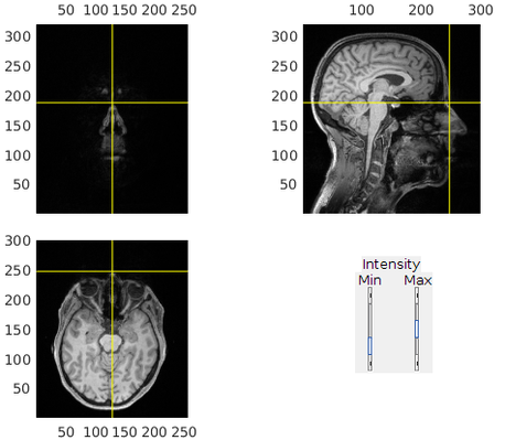
Left preauricular (LPA, press l)
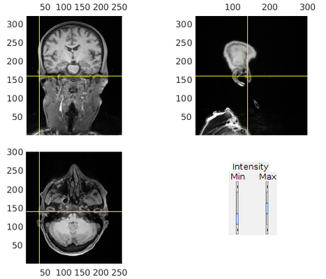
Right preauricular (RPA, press r)

3. Refine alignment with head shape points
ft_volumerealign
% headshape should be stored along with the MEG data
headshapef = '/path/to/MEGdata.fif';

cfg             = [];
cfg.method      = 'headshape';
headshape       = ft_read_headshape(headshapef);
headshape       = ft_convert_units(headshape, 'mm');
cfg.headshape.headshape = headshape;
cfg.coordsys    = 'neuromag';
cfg.headshape.interactive = 'no';
mri_aligned     = ft_volumerealign(cfg, mri_aligned);
This step can also be done interactively to further refine the alignment (set cfg.headshape.interactive = 'yes').
4. Create a head model (3D surfaces for brain, skull and scalp)
Segment and extract brain, skull and scalp surfaces.
ft_prepare_headmodel
cfg             = [];
cfg.output      = {'brain','skull','scalp'};
mri_seg         = ft_volumesegment(cfg, mri_aligned);

cfg             = [];
cfg.method      = 'singleshell';
cfg.tissue      = {'brain','skull','scalp'};
headmodel       = ft_prepare_headmodel(cfg,mri_seg);
Surfaces
Note that only the scalp surface is necessary to create the head model for source localization. It looks much nicer on the upcoming figures with 3 layers, though.
5. Load and align a template grid to the head model
ft_prepare_sourcemodel
cfg = [];
cfg.grid.warpmni   = 'yes';
cfg.grid.template  = template_grid;
cfg.grid.nonlinear = 'yes';
cfg.grid.unit      = 'mm';
cfg.mri            = mri_aligned;
grid               = ft_prepare_sourcemodel(cfg);

% save the results
save(fullfile(sujdir,'source_head_model.mat'), 'mri', 'mri_aligned','mri_seg','headmodel','grid','headshape');
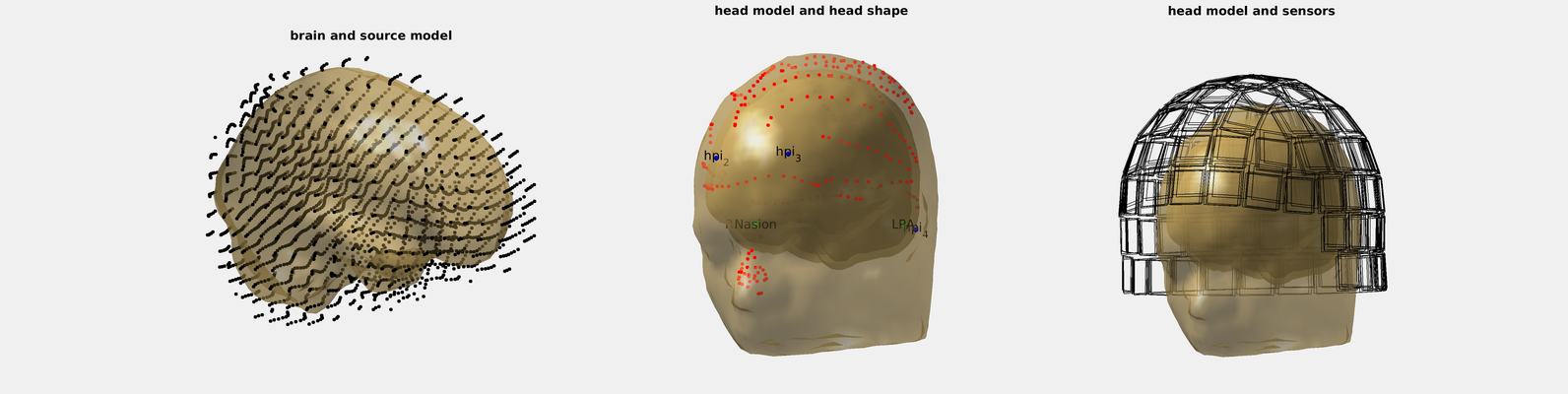
6. Plot the results and check
Check results
% make a figure with enough space for 3 panels horizontally
p = get(0,'defaultfigureposition'); p(3) = p(3) * 3;
figure('name',suj,'numbertitle','off','position',p);

% first panel with brain and aligned grid
subplot(131);
hm = headmodel;hm.bnd = hm.bnd(1);% use only the first layer (brain)
ft_plot_vol(hm,'vertexcolor','none','facecolor','skin','edgecolor','none','facealpha',.5);
ft_plot_mesh(grid.pos(grid.inside,:));%
lighting gouraud
material shiny
camlight
view(90,0)
rotate3d on
drawnow
title('brain and source model')

% second panel with head shape points, HPI coils, scalp, skull, and brain.
subplot(132);
ft_plot_vol(headmodel,'vertexcolor','none','facecolor','skin','edgecolor','none','facealpha',.5);
ft_plot_headshape(headshape);
hpi = find(cellfun(@(x)~isempty(x),regexp(headshape.label,'hpi_.*')));
hold on
scatter3(headshape.pos(hpi,1),headshape.pos(hpi,2),headshape.pos(hpi,3),200,'b','.');
text(headshape.pos(hpi,1),headshape.pos(hpi,2),headshape.pos(hpi,3), headshape.label(hpi), 'HorizontalAlignment', 'center', 'VerticalAlignment', 'middle', 'Interpreter', 'tex');
lighting gouraud
material shiny
camlight
view(-150,0)
rotate3d on
drawnow
title('head model and head shape')

% third panel with head and MEG sensors
subplot(133);cla
ft_plot_vol(headmodel,'vertexcolor','none','facecolor','skin','edgecolor','none','facealpha',.5);
lighting gouraud
material shiny
camlight
view(-180,0)
drawnow
% Below is the list of all runs by this subject
allruns = {'run01_tsss.fif','run02_tsss.fif','run03_tsss.fif'};
for i_run = 1:numel(allruns)
    h = ft_read_header(allruns{i_run});
    h.grad = ft_convert_units(h.grad, 'mm');

    ft_plot_sens(h.grad);
end
rotate3d on
drawnow
title('head model and sensors')
drawnow

% save as image and as .fig
export_fig(hfig,fullfile(rootdir,'figures','headmodel',['headmodel_' suj '.png']),'-nocrop','-r300'); % https://github.com/altmany/export_fig
saveas(hfig,fullfile(rootdir,'figures','headmodel',['headmodel_' suj '.fig']))
At this point we recommend to check (use the mouse to rotate the heads):
- The grid spans the whole brain and about a cm beyond
- The head shape points are aligned to the scalp surface
- Nasion, LPA and RPA are where they are supposed to be (although the head shape realignment moved them from where we clicked earlier)
- HPI coils are in place
- Sensors are where they are supposed to be (note that if you transformed the coordinate frame with Maxfilter to one recording run, the sensors should superimpose exactly, otherwise not)
Related articles
, multiple selections available, Use left or right arrow keys to navigate selected items